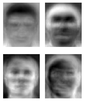
The above images are eigenfaces … which are statistically distilled basic components of human faces … from which ANY human face can be reconstructed as a combination of the above basic components. It’s a great mathematical trick – particularly if you’re into the whole mass surveillance and electronic police state thing.
If you are more into the whole, helping people and medical care thing, check out the global consortia at ENIGMA who have been carrying out massive genetic and brain scanning studies – like this one involving 437,607 SNPs in 31,622 voxels in 731 subjects using their new method, vGeneWAS, to study Alzheimer’s Disease:
“We hypothesized that vGeneWAS would, in some situations, have greater power to detect associations than existing SNP-based methods. One such situation might be when a gene contains many loci with weak individual effects. In addition, we expected that vGeneWAS would have greater overall power than mass SNP-based methods, like vGWAS, because of the drastic reduction in the effective number of statistical tests performed.”
The vGeneWAS method relies on the calculation of “eigenSNPs” which are eigenvectors that describe a matrix of n subjects by m SNPs in an individual gene (an n-x-m matrix of 1’s,0’s,-1’s for aa, aA, AA genotypes). EigenSNPs are sort of like eigenfaces insofar as eigenSNPs (which are not actual SNPs) capture the majority of variance, or the basic essence of an individual gene … but seriously, you should read the original article ’cause every stats test I ever took totally punched me in the face.
In any case, the eigenSNP-by-voxel method pulled out some legit results such as rs2373115 (where the G-allele confers risk) in the GAB2 gene which has repeatedly been implicated in the risk of age-related late-onset Alzheimer’s Disease (in folks who carry ApoE4 rs429358(C) alleles). The authors found that the genetic risk of AD conferred by GAB2 may arise by way of GAB2’s effect on brain structure in the periventricular areas, which have been known to be among the first brain regions to show AD-related changes (time-lapse movie of AD tissue loss in the brain).


Leave a comment