Feeds:
Posts
Comments

Posts Tagged ‘Mental disorder’

BeFunky_OldPhoto_12

Have you ever read the DSM and thought you had EVERYTHING? Me too.

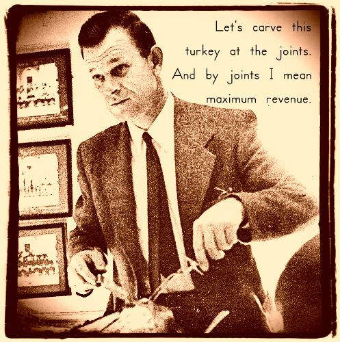
And that, sort of,  has always been a big problem … that it is really hard to separate the normal experience of anguish and suffering as part of our everyday mental and emotional lives from what is labelled a “disorder”. At the same time, however, patients, doctors and payors need some type of common reference so as to keep the diagnosis and treatment of mental suffering in-line with the way other medical illnesses are handled. So, everyone (in psychiatry, at least) knows the DSM will always be highly flawed and yet also highly necessary … so, you know, just try and live with it … but don’t expect, for a moment, to search for and find discrete genetic variants that correspond to DSM categories of mental disorders. No … because the DSM categories do not correspond well to the underlying biology of the CNS … the DSM does not “cut nature at its joints” so to speak.

Russ Poldrack provides a glimpse into what the future of diagnosing mental illness might look like using slightly more objective, quantifiable and biologically relevant measures of the brain’s physiological processes.

Also, I stumbled onto an awesome read about the creation of DSM-5 entitled, The Book of Woe

The overall thrust of the manual [DSM-5], the BPS complained, was to identify the source of psychological suffering “as located within individuals” rather than in their “relational context,” and to overlook the “undeniable social causation of many such problems.”  The APA could hardly deny any of this. As Regier had told the consumer groups on the conference call, the manual’s new organizational structure was designed to reflect “what we’ve learned about the brain, behavior, and genetics during the past two decades.” It doesn’t get much more “within the individual” and outside the “relational context” than that. (p. 239)

“Dereification is just as dumb as reinfication,” he [Allen Frances] told me. “A construct is just a construct – not to be worshiped and not to be denigrated.” Psychiatry, he was saying, has to live in the tension between the desire for certainty about the nature of our suffering and the impossibility of understanding it (or ourselves) completely. A DSM that tries to end this tension by turning itself into a living document was bound to collapse into chaos; that was the cardinal error of the incompetent DSM-5 regime. (p. 279)

“What [Dr. Thomas] Insel [Director of NIMH] heard “over and over again” on his tour was that psychiatrists were tired of being trapped by the DSM. “We are so embedded in this structure,” he told me. He and his colleagues had spent so much time diagnosing mental disorders that “we actually believe they are real. But there’s no reality. These are just constructs. There’s no reality to schizophrenia and depression.” Indeed, Insel said, “we might have to stop using terms like depression and schizophrenia, because they are getting in our way, confusing things.” Thirty years after Spitzer burned down DSM-II and built the DSM-III in its ashes, psychiatry might once again have to “just sort of start over.”” (p.340)

Yikes! after reading The Book of Woe, DSM-5 sounds, um, totally wack … if not a tool flagrantly designed to further commodify human suffering for the benefit of a medico-industrial complex. NIMH Director Thomas Insel’s recent announcement that, “NIMH will be re-orienting its research away from DSM categories.” suggests a future where diagnosis will based on biological measures and treatments are directed toward specific circuits.

Treatment for specific circuit dynamics sounds very promising. However, I thought Dr. Allen Frances, as quoted in The Book of Woe made a great point (p.346) that, “The trick is to develop a healing relationship, to care for the person not just the disorder, to diagnose and treat cautiously, and to see the healthy part of the person not just the sick.”

* Maybe that is the hope of this blog also … to take out and explore the intricate biological & molecular parts … but also to try and place them back into their original evolutionary, living, breathing, copulating (or more often the case of just thinking about copulating) “whole” human being.

Read Full Post »

Mitochondrial damage is associated with premature aging in the body and related disorders such as Parkinson’s Disease in the brain.  If you want to grow old and healthy … be nice to your mitochondria … eat healthy foods and exercise.

When mitochondria are damaged, cells can use proteolysis to clean them out, but when this cleaning out process fails … trouble ensues.   PINK1 plays a role on the clearance of damaged mitochondria as revealed by Dr. Derek P. Narendra and colleagues: PINK1 Is Selectively Stabilized on Impaired Mitochondria to Activate Parkin

Since neurons in the Substantia Nigra are postmitotic, any mitochondrial damage they acquire could accumulate over an organism’s lifetime, leading to progressive mitochondrial dysfunction—including increased oxidative stress, decreased calcium buffering capacity, loss of ATP, and, eventually, cell death—unless quality control processes eliminate the damaged mitochondria.

The findings we report in this paper suggest a new model in which PINK1 and Parkin together sense mitochondria in distress and selectively target them for degradation. In this pathway, PINK1 acts as a flag that accumulates on dysfunctional mitochondria and then signals to Parkin, which tags these mitochondria for destruction. Since disease-causing mutations in PINK1 or Parkin disrupt this pathway, patients with these mutations may not be able to clean up their damaged mitochondria, leading to the neuronal damage typical of parkinsonism.

Dr. Terry Wahls has some very inspiring experiences to share on the topic of mitochondrial care.

Read Full Post »

Corticotropin-releasing hormone
Image via Wikipedia

According to the authors of  “Protective effect of CRHR1 gene variants on the development of adult depression following childhood maltreatment: replication and extension”  [PMID: 19736354], theirs is “the first instance of Genes x Environment research that stress has been ascertained by more than 1 study using the same instrument“.  The gene they speak of is the Corticotropin-releasing hormone receptor 1 (CRHR1) gene (SNPs rs7209436, rs110402, rs242924 which can form a so-called T-A-T haplotype which has been associated with protection from early life stress (as ascertained using the Childhood Trauma Questionnaire CTQ)).

The research team examined several populations of adults and, like many other studies, found that early life stress was associated with symptoms of depressive illness but, like only 1 previous study, found that the more T-A-T haplotypes a person has (0,1,or 2) the less likely they were to suffer these symptoms.

Indeed, the CRHR1 gene is an important player in a complex network of hormonal signals that regulate the way the body (specifically the hypothalamic pituitary adrenal axis) transduces the effects of stress.  So it seems quite reasonable to see that individual differences in ones ability to cope with stress might correlate with genotype here.   The replication seems like a major step forward in the ongoing paradigm shift from “genes as independent risk factors” to “genetic risk factors being dependent on certain environmental forces”.  The authors suggest that a the protective T-A-T haplotype might play a role in the consolidation of emotional memories and that CRHR1 T-A-T carriers might have a somewhat less-efficient emotional memory consolidation (sort of preventing disturbing memories from making it into long-term storage in the first place?) – which is a very intriguing and testable hypothesis.

On a more speculative note … consider the way in which the stress responsivity of a developing child is tied to its mother’s own stress responsivity.  Mom’s own secretion of CRH from the placenta is known to regulate gestational duration and thus the size, heartiness and stress responsiveness of her newborn.  The genetic variations are just passed along from generation to generation and provide some protection here and there in an intertwined cycle of life.

The flowers think they gave birth to seeds,
The shoots, they gave birth to the flowers,
And the plants, they gave birth to the shoots,
So do the seeds they gave birth to plants.
You think you gave birth to the child.
None thinks they are only entrances
For the life force that passes through.
A life is not born, it passes through.

anees akbar

Reblog this post [with Zemanta]

Read Full Post »

Twin studies have long suggested that genetic variation is a part of healthy and disordered mental life.  The problem however – some 10 years now since the full genome sequence era began – has been finding the actual genes that account for this heritability.

It sounds simple on paper – just collect lots of folks with disorder X and look at their genomes in reference to a demographically matched healthy control population.  Voila! whatever is different is a candidate for genetic risk.  Apparently, not so.

The missing heritability problem that clouds the birth of the personal genomes era refers to the baffling inability to find enough common genetic variants that can account for the genetic risk of an illness or disorder.

There are any number of reasons for this … (i) even as any given MZ and DZ twin pair shares genetic variants that predispose them toward the similar brains and mental states, it may be the case that different MZ and DZ pairs have different types of rare genetic variation thus diluting out any similar patterns of variation when large pools of cases and controls are compared …  (ii) also, the way that the environment interacts with common risk-promoting genetic variation may be quite different from person to person – making it hard to find variation that is similarly risk-promoting in large pools of cases and controls … and many others I’m sure.

One research group recently asked whether the type of common genetic variation(SNP vs. CNV) might inform the search for the missing heritability.  The authors of the recent paper, “Genome-wide association study of CNVs in 16,000 cases of eight common diseases and 3,000 shared controls” [doi:10.1038/nature08979] looked at an alternative to the usual SNP markers – so called common copy number variants (CNVs) – and asked if these markers might provide a stronger accounting for genetic risk.  While a number of previous papers in the mental health field have indeed shown associations with CNVs, this massive study (some 3,432 CNV probes in 2000 or so cases and 3000 controls) did not reveal an association with bipolar disorder.  Furthermore, the team reports that common CNV variants are already in fairly strong linkage disequilibrium with common SNPs and so perhaps may not have reached any farther into the abyss of rare genetic variation than previous GWAS studies.

Disappointing perhaps, but a big step forward nonetheless!  What will the personal genomes era look like if we all have different forms of rare genetic variation?

Reblog this post [with Zemanta]

Read Full Post »

According to wikipedia, “Jean Philippe Arthur Dubuffet (July 31, 1901 – May 12, 1985) was one of the most famous French painters and sculptors of the second half of the 20th century.”  “He coined the term Art Brut (meaning “raw art,” often times referred to as ‘outsider art’) for art produced by non-professionals working outside aesthetic norms, such as art by psychiatric patients, prisoners, and children.”  From this interest, he amassed the Collection de l’Art Brut, a sizable collection of artwork, of which more than half, was painted by artists with schizophrenia.  One such painting that typifies this style is shown here, entitled, General view of the island Neveranger (1911) by Adolf Wolfe, a psychiatric patient.

Obviously, Wolfe was a gifted artist, despite whatever psychiatric diagnosis was suggested at the time.  Nevertheless, clinical psychiatrists might be quick to point out that such work reflects the presence of an underlying thought disorder (loss of abstraction ability, tangentiality, loose associations, derailment, thought blocking, overinclusive thinking, etc., etc.) – despite the undeniable aesthetic beauty in the work.  As an ardent fan of such art,  it made me wonder just how “well ordered” my own thoughts might be.  Given to being rather forgetful and distractable, I suspect my thinking process is just sufficiently well ordered to perform the routine tasks of day-to-day living, but perhaps not a whole lot more so.  Is this bad or good?  Who knows.

However, Krug et al., in their recent paper, “The effect of Neuregulin 1 on neural correlates of episodic memory encoding and retrieval” [doi:10.1016/j.neuroimage.2009.12.062] do note that the brains of unaffected relatives of persons with mental illness show subtle differences in various patterns of activation.  It seems that when individuals are using their brains to encode information for memory storage, unaffected relatives show greater activation in areas of the frontal cortex compared to unrelated subjects.  This so-called encoding process during episodic memory is very important for a healthy memory system and its dysfunction is correlated with thought disorders and other aspects of cognitive dysfunction.  Krug et al., proceed to explore this encoding process further and ask if a well-known schizophrenia risk variant (rs35753505 C vs. T) in the neuregulin-1 gene might underlie this phenomenon.  To do this, they asked 34 TT, 32 TC and 28 CC individuals to perform a memory (of faces) game whilst laying in an MRI scanner.

The team reports that there were indeed differences in brain activity during both the encoding (storage) and retrieval (recall) portions of the task – that were both correlated with genotype – and also in which the CC risk genotype was correlated with more (hyper-) activation.  Some of the brain areas that were hyperactivated during encoding and associated with CC genotype were the left middle frontal gyrus (BA 9), the bilateral fusiform gyrus and the left middle occipital gyrus (BA 19).  The left middle occipital gyrus showed gene associated-hyperactivation during recall.  So it seems, that healthy individuals can carry risk for mental illness and that their brains may actually function slightly differently.

As an ardent fan of Art Brut, I confess I hoped I would carry the CC genotype, but alas, my 23andme profile shows a boring TT genotype.  No wonder my artwork sucks.  More on NRG1 here.

Reblog this post [with Zemanta]

Read Full Post »

wotd044
Image by theloushe via Flickr

** PODCAST accompanies this post**

I have a little boy who loves to run and jump and scream and shout – a lot.  And by this, I mean running – at full speed and smashing his head into my gut,  jumping – off the couch onto my head,  screaming – spontaneous curses and R-rated body parts and bodily functions.  I hope you get the idea.  Is this normal? or (as I oft imagine) will I soon be sitting across the desk from a school psychologist pitching me the merits of an ADHD diagnosis and medication?

Of course, when it comes to behavior, there is not a distinct line one can cross from normal to abnormal.  Human behavior is complex, multi-dimensional and greatly interpreted through the lens of culture.  Our present culture is highly saturated by mass-marketing, making it easy to distort a person’s sense of “what’s normal” and create demand for consumer products that folks don’t really need (eg. psychiatric diagnoses? medications?).   Anyhow, its tough to know what’s normal.  This is an important issue to consider for those (mass-marketing hucksters?) who might be inclined to promote genetic data as “hard evidence” for illness, disorder or abnormality of some sort.

With this in mind, I really enjoyed a recent paper by Stollstorff et al., “Neural response to working memory load varies by dopamine transporter genotype in children” [doi:10.1016/j.neuroimage.2009.12.104] who asked how the brains of healthy children functioned, even though they carry a genotype that has been widely associated with the risk of ADHD.  Healthy children who carry genetic risk for ADHD. Hmm, might this be my boy?

The researchers looked at a 9- vs. 10-repeat VNTR polymorphism in the 3′-UTR of the dopamine transporter gene (DAT1).  This gene – which encodes the very protein that is targeted by so many ADHD medications – influences the re-uptake of dopamine from the synaptic cleft.  In the case of 10/10 genotypes, it seems that DAT1 is more highly expressed, thus leading to more re-uptake and hence less dopamine in the synaptic cleft.  Generally, dopamine is needed to enhance the signal/noise of neurotransmission, so – at the end of the day – the 10/10 genotype is considered less optimal than the 9/9-repeat genotype.  As noted by the researchers, the ADHD literature shows that the 10-repeat allele, not the 9-repeat, is most often associated with ADHD.

The research team asked these healthy children (typically developing children between 7 and 12 years of age) to perform a so-called N-back task which requires that children remember words that are presented to them one-at-a-time.  Each time a new word is presented, the children had to decide whether that word was the same as the previous word (1-back) or the previous, previous word (2-back).  Its a maddening task and places an extreme demand on neural circuits involved in active maintenance of information (frontal cortex) as well as inhibition of irrelevant information that occurs during updating (basal ganglia circuits).

As the DAT1 protein is widely expressed in the basal ganglia, the research team asked where in the brain was variation in the DAT1 (9- vs. 10-repeat) associated with neural activity?  and where was there a further difference between 1-back and 2-back?  Indeed, the team finds that brain activity in many regions of the basal ganglia (caudate, putamen, substantia nigra & subthalamic nucleus) were associated with genetic variation in DAT1.  Neat!  the gene may be exerting an influence on brain function (and behavior) in healthy children, even though they do not carry a diagnosis.  Certainly, genes are not destiny, even though they do influence brain and behavior.

What was cooler to me though, is the way the investigators examined the role of genetic variation in the 1-back (easy or low load condition) vs. 2-back (harder, high-load condition) tasks.  Their data shows that there was less of an effect of genotype on brain activation in the easy tasks.  Rather, only when the task was hard, did it become clear that the basal ganglia in the 10/10 carriers was lacking the necessary brain activation needed to perform the more difficult task.  Thus, the investigators reveal that the genetic risk may not be immediately apparent under conditions where heavy “loads” or demands are not placed on the brain.  Cognitive load matters when interpreting genetic data!

This result made me think that genes in the brain might be a lot like genes in muscles.  Individual differences in muscle strength are not associated with genotype when kids are lifting feathers.  Only when kids are actually training and using their muscles, might one start to see that some genetically advantaged kids have muscles that strengthen faster than others.  Does this mean there is a “weak muscle gene” – yes, perhaps.  But with the proper training regimen, children carrying such a “weak muscle gene” would be able to gain plenty of strength.

I guess its off to the mental and physical gyms for me and my son.

** PODCAST accompanies this post** also, here’s a link to the Vaidya lab!

Reblog this post [with Zemanta]

Read Full Post »

Recreated :File:Neuron-no labels2.png in Inksc...
Image via Wikipedia

The A-to-T SNP rs7794745 in the CNTNAP2 gene was found to be associated with increased risk of autism (see Arking et al., 2008).  Specifically, the TT genotype, found in about 15% of individuals, increases these folks’ risk by about 1.2-1.7-fold.  Sure enough, when I checked my 23andMe profile, I found that I’m one of these TT risk-bearing individuals.  Interesting, although not alarming since me and my kids are beyond the age where one typically worries about autism.  Still, one can wonder if such a risk factor might have exerted some influence on the development of my brain?

The recent paper by Tan et al., “Normal variation in fronto-occipital circuitry and cerebellar structure with an autism-associated polymorphism of CNTNAP2” [doi:10.1016/j.neuroimage.2010.02.018 ] suggests there may be subtle, but still profound influences of the TT genotype on brain development in healthy individuals.  According to the authors, “homozygotes for the risk allele showed significant reductions in grey and white matter volume and fractional anisotropy in several regions that have already been implicated in ASD, including the cerebellum, fusiform gyrus, occipital and frontal cortices. Male homozygotes for the risk alleles showed greater reductions in grey matter in the right frontal pole and in FA in the right rostral fronto-occipital fasciculus compared to their female counterparts who showed greater reductions in FA of the anterior thalamic radiation.”

The FA (fractional anisotropy – a measurement of white-matter or myelination) results are consistent with a role of CNTNAP2 in the establishment of synaptic contacts and other cell-cell contacts especially at Nodes of Ranvier – which are critical for proper function of white-matter tracts that support rapid, long-range neural transmission.  Indeed, more severe mutations in CNTNAP2  have been associated with cortical dysplasia and focal epilepsy (Strauss et al., 2006).

Subtle changes perhaps influencing long-range information flow in my brain – wow!

More on CNTNAP2 … its evolutionary history and role in language development.

Reblog this post [with Zemanta]

Read Full Post »

If you’re a coffee drinker, you may have noticed the new super-sized portions available at Starbucks.  On this note, it may be worth noting that caffeine is a potent psychoactive substance of which – too much – can turn your buzz into a full-blown panic disorder.  The Diagnostic and Statistical Manual for psychiatry outlines a number of caffeine-related conditions mostly involving anxieties that can arise when the natural alertness-promoting effects are pushed to extremes.  Some researchers have begun to explore the way the genome interacts with caffeine and it is likely that many genetic markers will surface to explain some of the individual differences in caffeine tolerance.

Here’s a great paper, “Association between ADORA2A and DRD2 Polymorphisms and Caffeine-Induced Anxiety” [doi: 10.1038/npp.2008.17] wherein polymorphisms in the adenosine A2A receptor (ADORA2A encodes the protein that caffeine binds to and antagonizes) – as well as the dopamine D2 receptor (DRD2 encodes a protein whose downstream signals are normally counteracted by A2A receptors) — show associations with anxiety after the consumption of 150mg of caffeine (about an average cup of coffee – much less than the super-size, super-rich cups that Starbucks sells).  The variants, rs5751876 (T-allele), rs2298383 (T-allele) and rs4822492 (G-allele) from the ADORA2A gene as well as rs1110976 (-/G genotype) from the DRD2 gene showed significant increases in anxiety in a test population of 102 otherwise-healthy light-moderate regular coffee drinkers.

My own 23andMe data only provides a drop of information suggesting I’m protected from the anxiety-promoting effects.  Nevertheless, I’ll avoid the super-sizes.
rs5751876 (T-allele)  C/C – less anxiety
rs2298383 (T-allele) – not covered
rs4822492 (G-allele) – not covered
rs1110976 (-/G genotype) – not covered

Reblog this post [with Zemanta]

Read Full Post »

03230052.JPG
Image by mbrownstone via Flickr

Walter Dean Myers, an author of The Young Landlords and many other classic coming of age novels once remarked, “The special place of the young adult novel should be in its ability to address the needs of the reader to understand his or her relationships with the world, with each other, and with adults.”  Indeed, the wonderful elaborations of psychosocial development that occur during the teenage years makes for a vivid and tumultuous time – worthy of many a book – especially those like Myers’ that so help adolescents to cope.  During this time, a child’s brain and body is supplanted by adult systems, which, from a physiological point of view, place the adolescent’s mind and body at the mercy of thousands of shifting biochemical processes.  Such a notion of the shifting sands of adolescence were brought to mind while reading a research article focused on one – just one single example – of biochemical change.

The paper entitled, “Cortico-striatal synaptic defects and OCD-like behaviors in SAPAP3 mutant mice” [doi: 10.1038/nature06104] points out that mice who lack the function of the post-synaptic density scaffolding protein encoded by the SAPAP3 gene display excessive grooming and other behaviors reminiscent of obsessive compulsive disorder – a condition that frequently emerges during adolescence.  One of the main findings of the paper is that a normal developmental shift of NR2B –> NR2A subunits of the NMDA receptor does NOT seem to occur – rendering the SAPAP3 mutant mice with an immature form of NMDA receptor.  The authors suggest that this may be the underlying reason for the aberrant behavior, and were able to normalize the mutant mice by re-introducing SAPAP3 protein via a lentiviral-mediated expression vector placed in the striatum.

Gosh.  This NR2B –> NR2A shift is just one example – one grain – in the shifting biochemical sands of development.  Just one of thousands.  How did my brain ever make it through?

Reblog this post [with Zemanta]

Read Full Post »

Summer, Brody and Audric Hug
Image by cobalt123 via Flickr

If you have a minute, check out this “Autism Sensory Overload Simulation” video to get a feel for the perceptual difficulties experienced by people with autism spectrum disorders.  A recent article, “Critical Period Plasticity Is Disrupted in the Barrel Cortex of Fmr1 Knockout Mice” [doi: 10.1016/j.neuron.2010.01.024] provides some clues to the cellular mechanisms that are involved in this phenomenon.  The authors examined the developing somatosensory cortex in lab mice who carry a mutation in a gene called FMR1.  The normal function of this gene is to help synapses mature and optimize their strength through a process known as activity-dependent plasticity.  This a kind of “use-it-or-lose-it” neural activity that is important when you are practicing and practicing to learn something new – say, like riding a bike, or learning a new language.  Improvements in performance that come from “using” the circuits in the brain are correlated with optimized synaptic connections – via a complex set of biochemical reactions (eg. AMPA receptor trafficking).

When FMR1 is not functioning, neuronal connections (in this case, synapses that connect the thalamus to the somatosensory cortex) cannot mature and develop properly.  This wreaks havoc in the developing brain where maturation can occur in successive critical periods – where the maturation of one circuit is needed to ensure the subsequent development of another.  Hence, the authors suggest, the type of sensory overload reported in the autism spectrum disorders may be related to a similar type of developmental anomaly in the somatosensory cortex.

Reblog this post [with Zemanta]

Read Full Post »

Diagram to illustrate Minute Structure of the ...
Image via Wikipedia

For a great many reasons, research on mental illness is focused on the frontal cortex.  Its just a small part of the brain, and certainly, many things can go wrong in other places during brain/cognitive development, but, it remains a robust finding, that when the frontal cortex is not working well, individuals have difficulties in regulating thoughts and emotions.  Life is difficult enough to manage, let alone without a well functioning frontal cortex.  So its no surprise that many laboratories look very closely at how this region develops prenatally and during childhood.

One of the more powerful genetic methods is the analysis of gene expression via microarrays (here is a link to a tutorial on this technology).  When this technology is coupled with extremely careful histological analysis and dissection of cortical circuits in the frontal cortex, it begins to become possible to begin to link changes in gene expression with the physiological properties of specific cells and local circuits in the frontal cortex. The reason this is an exciting pursuit is because the mammalian neocortex is organized in a type of layered fashion wherein 6 major layers have different types of connectivity and functionality.  The developmental origins of this functional specificity are thought to lie in a process known as radial migration (here is a video of a neuron as it migrates radially and finds its place in the cortical hierarchy).  As cells are queued out of the ventricular zone, and begin their migration to the cortical surface, they are exposed to all sorts of growth factors and morphogens that help them differentiate and form the proper connectivities.  Thus, the genes that regulate this process are of keen interest to understanding normal and abnormal cognitive development.

Here’s an amazing example of this – 2 papers entitled, “Infragranular gene expression disturbances in the prefrontal cortex in schizophrenia: Signature of altered neural development?” [doi:10.1016/j.nbd.2009.12.013] and “Molecular markers distinguishing supragranular and infragranular layers in the human prefrontal cortex [doi:10.1111/j.1460-9568.2007.05396.x] both by Dominique Arion and colleagues.  In both papers, the authors ask, “what genes are differentially expressed in different layers of the cortex?”.  This is a powerful line of inquiry since the different layers of cortex are functionally different in terms of their connectivity.  For example, layers II-III (the so-called supragranular layers) are known to connect mainly to other cortical neurons – which is different functionally than layers V-VI (the so-called infragranular layers) that connect mainly to the striatum (layer V) and thalamus (layer VI).  Thus, if there are genes whose expression is unique to a layer, then one has a clue as to how that gene might contribute to normal/abnormal information processing.

The authors hail from a laboratory that is well-known for work over many years on fine-scaled histological analysis of the frontal cortex at the University of Pittsburgh and used a method called, laser capture microdissection, where post-mortem sections of human frontal cortex (area 46) were cut to separate the infragraular layer from the supragranular layer.  The mRNA from these tissue sections was then used for DNA microarray hybridization.  Various controls, replicate startegies and in-situ tissue hybridizations were then employed to validate the initial microarray results.

In first paper, the where the authors compare infra vs. supragranular layers, they report that 40 genes were more highly expressed in the supragranular layers (HOP, CUTL2 and MPPE1 were among the most enriched) and 29 genes were highly expressed in the infragranular layers (ZNF312, CHN2, HS3ST2 were among the most enriched).  Other differentially expressed genes included several that have previously been implicated in cortical layer formation such as RLN, TLX-NR2E1, SEMA3E, PCP4, SERPINE2, NR2F2/ARP1, PCDH8, WIF1, JAG1, MBP.  Amazing!! A handful of genes that seem to label subpopulations of projection neurons in the frontal cortex.  Polymorphic markers for these genes would surely be powerful tools for imaging-genetic studies on cognitive development.

In the second paper, the authors compare infra vs. supragranular gene expression in post-mortem brains from patients with schizophrenia and healthy matched controls. Using the same methods, the team reports both supra- and infragranular gene expression changes in schizophrenia (400 & 1200 differences respectively) – more than 70% of the differences appearing to be reductions in gene expression in schizophrenia. Interestingly, the team reports that the genes that were differentially expressed in the infragranular layers provided sufficient information to discriminate between cases and controls, whilst the gene expression differences in the supragranular layers did not.  More to the point, the team finds that 51 genes that were differentially expressed in infra- vs. supragranular expression were also differentially expressed in cases vs. controls  (many of these are also found to be associated in population genetic association studies of schiz vs. control as well!).  Thus, the team has identified layer (function) -specific genes that are associated with schizophrenia.  These genes, the ones enriched in the infragranular layers especially, seem to be at the crux of a poorly functioning frontal cortex.

The authors point to 3 such genes (SEMA3E, SEMA6D, SEMA3C) who happen to members of the same gene family – the semaphorin gene family.  This gene family is very important for the neuronal guidance (during radial migration), morphology, pruning and other processes where cell shape and position are regulated.  The authors propose that the semaphorins might act as “integrators” of various forms of wiring during development and in adulthood.  More broadly, the authors provide a framework to understand how the development of connectivity on the frontal cortex is regulated by genetic factors – indeed, many suspected genetic risk factors play a role in the developmental pathways the authors have focused on.

Reblog this post [with Zemanta]

Read Full Post »

One of the complexities in beginning to understand how genetic variation relates to cognitive function and behavior is that – unfortunately – there is no gene for “personality”, “anxiety”, “memory” or any other type of “this” or “that” trait.  Most genes are expressed rather broadly across the entire brain’s cortical layers and subcortical systems.  So, just as there is no single brain region for “personality”, “anxiety”, “memory” or any other type of “this” or “that” trait, there can be no such gene.  In order for us to begin to understand how to interpret our genetic make-up, we must learn how to interpret genetic variation via its effects on cells and synapses – that go on to function in circuits and networks.  Easier said than done?  Yes, but perhaps not so intractable.

Here’s an example.  One of the most well studied circuits/networks/systems in the field of cognitive science are so-called basal-ganglia-thalamcortical loops.  These loops have been implicated in a great many forms of cognitive function involving the regulation of everything from movement, emotion and memory to reasoning ability.  Not surprisingly, neuroimaging studies on cognitive function almost always find activations in this circuitry.  In many cases, the data from neuroimaging and other methodologies suggests that one portion of this circuitry – the frontal cortex – plays a role in the representation of such aspects as task rules, relationships between task variables and associations between possible choices and outcomes.  This would be sort of like the “thinking” part of our mental life where we ruminate on all the possible choices we have and the ins and outs of what each choice has to offer.  Have you ever gone into a Burger King and – even though you’ve known for 20 years what’s on the menu – you freeze up and become lost in thought just as its your turn to place your order?  Your frontal cortex is at work!

The other aspect of this circuitry is the subcortical basla ganglia, which seems to play the downstream role of processing all that ruminating activity going on in the frontal cortex and filtering it down into a single action.  This is a simple fact of life – that we can be thinking about dozens of things at a time, but we can only DO 1 thing at a time.  Alas, we must choose something at Burger King and place our order.  Indeed, one of the hallmarks of mental illness seems to be that this circuitry functions poorly – which may be why individuals have difficulty in keeping their thoughts and actions straight – the thinking clearly and acting clearly aspect of healthy mental life.  Certainly, in neurological disorders such as Parkinson’s Disease and Huntington’s Disease, where this circuitry is damaged, the ability to think and move one’s body in a coordinated fashion is disrupted.

Thus, there are at least 2 main components to a complex system/circuits/networks that are involved in many aspects of learning and decision making in everyday life.  Therefore, if we wanted to understand how a gene – that is expressed in both portions of this circuitry – inflenced our mental life, we would have to interpret its function in relation to each specific portion of the circuitry.  In otherwords, the gene might effect the prefrontal (thinking) circuitry in one way and the basla-ganglia (action-selection) circuitry in a different way.  Since we’re all familiar with the experience of walking in to a Burger King and seeing folks perplexed and frozen as they stare at the menu, perhaps its not too difficult to imagine that a gene might differentially influence the ruminating process (hmm, what shall I have today?) and the action selection (I’ll take the #3 combo) aspect of this eveyday occurrance (for me, usually 2 times per week).

Nice idea you say, but does the idea flow from solid science?  Well, check out the recent paper from Cindy M. de Frias and colleagues “Influence of COMT Gene Polymorphism on fMRI-assessed Sustained and Transient Activity during a Working Memory Task.” [PMID: 19642882].  In this paper, the authors probed the function of a single genetic variant (rs4680 is the Methionine/Valine variant of the dopamine metabolizing COMT gene) on cognitive functions that preferentially rely on the prefronal cortex as well as mental operations that rely heavily on the basal-ganglia.  As an added bonus, the team also probed the function of the hippocampus – yet a different set of circuits/networks that are important for healthy mental function.  OK, so here is 1 gene who is functioning  within 3 separable (yet connected) neural networks!

The team focused on a well-studied Methionine/Valine variant of the dopamine metabolizing COMT gene which is broadly expessed across the pre-frontal (thinking) part of the circuitry and the basal-ganglia part of the circuitry (action-selection) as well as the hippocampus.  The team performed a neuroimaging study wherein participants (11 Met/Met and 11 Val/Val) subjects had to view a series of words presented one-at-a-time and respond if they recalled that a word was a match to the word presented 2-trials beforehand  (a so-called “n-back task“).  In this task, each of the 3 networks/circuits (frontal cortex, basal-ganglia and hippocampus) are doing somewhat different computations – and have different needs for dopamine (hence COMT may be doing different things in each network).  In the prefrontal cortex, according to a theory proposed by Robert Bilder and colleagues [doi:10.1038/sj.npp.1300542] the need is for long temporal windows of sustained neuronal firing – known as tonic firing (neuronal correlate with trying to “keep in mind” all the different words that you are seeing).  The authors predicted that under conditions of tonic activity in the frontal cortex, dopamine release promotes extended tonic firing and that Met/Met individuals should produce enhanced tonic activity.  Indeed, when the authors looked at their data and asked, “where in the brain do we see COMT gene associations with extended firing? they found such associations in the frontal cortex (frontal gyrus and cingulate cortex)!

Down below, in the subcortical networks, a differerent type of cognitive operation is taking place.  Here the cells/circuits are involved in the action selection (press a button) of whether the word is a match and in the working memory updating of each new word.  Instead of prolonged, sustained “tonic” neuronal firing, the cells rely on fast, transient “phasic” bursts of activity.  Here, the modulatory role of dopamine is expected to be different and the Bilder et al. theory predicts that COMT Val/Val individuals would be more efficient at modulating the fast, transient form of cell firing required here.   Similarly, when the research team explored their genotype and brain activity data and asked, “where in the brain do we see COMT gene associations with transient firing? they found such associations in the right hippocampus.

Thus, what can someone who carries the Met/Met genotype at rs4680 say to their fellow Val/Val lunch-mate next time they visit a Burger King?  “I have the gene for obesity? or impulsivity? or “this” or “that”?  Perhaps not.  The gene influences different parts of each person’s neural networks in different ways.  The Met/Met having the advantage in pondering (perhaps more prone to annoyingly gaze at the menu forever) whist the Val/Val has the advantage in the action selecting (perhaps ordering promptly but not getting the best burger and fries combo).

Reblog this post [with Zemanta]

Read Full Post »

We hope, that you choke, that you choke.
Image by Corrie… via Flickr

Coping with fear and anxiety is difficult.  At times when one’s life, livelihood or loved one’s are threatened, we naturally hightenen our senses and allocate our emotional and physical resources for conflict.  At times, when all is well, and resources, relationships and relaxation time are plentiful, we should unwind and and enjoy the moment.  But most of us don’t.  Our prized cognitive abilities to remember, relive and ruminate on the bad stuff out there are just too well developed – and we suffer – some more than others  (see Robert Saplosky’s book “Why Zebras Don’t Get Ulcers” and related video lecture (hint – they don’t get ulcers because they don’t have the cognitive ability to ruminate on past events).  Such may be the flip side to our (homo sapiens) super-duper cognitive abilities.

Nevertheless, we try to understand our fears and axieties and understand their bio-social-psychological bases. A recent paper entitled, “A Genetically Informed Study of the Association Between Childhood Separation Anxiety, Sensitivity to CO2, Panic Disorder, and the Effect of Childhood Parental Loss” by Battaglia et al. [Arch Gen Psychiatry. 2009;66(1):64-71] brought to mind many of the complexities in beginning to understand the way in which some individuals come to suffer more emotional anguish than others.  The research team addressed a set of emotional difficulties that have been categorized by psychiatrists as “panic disorder” and involving sudden attacks of fear, sweating, racing heart, shortness of breath, etc. which can begin to occur in early adulthood.

Right off the bat, it seems that one of the difficulties in understanding such an emotional state(s) are the conventions (important for $$ billing purposes) used to describe the relationship between “healthy” and “illness” or “disorder”.  I mean, honestly, who hasn’t experienced what could be described as a mild panic disorder once or twice?  I have, but perhaps that doesn’t amount to a disorder.  A good read on the conflation of normal stress responses and disordered mental states is “Transforming Normality into Pathology: The DSM and the Outcomes of Stressful Social Arrangements” by Allan V. Horwitz.

Another difficulty in understanding how and why someone might experience such a condition has to do with the complexities of their childhood experience (not to mention genes). Child development and mental health are inextrictably related, yet, the relationship is hard to understand.  Certainly, the function of the adult brain is the product of countless developmental unfoldings that build upon one another, and certainly there is ample evidence that when healthy development is disrupted in a social or physical way, the consequences can be very unfortunate and long-lasting. Yet, our ability to make sense of how and why an individual is having mental and/or emotional difficulty is limited.  Its a complex, interactive and emergent set of processes.

What I liked about the Battaglia et al., article was the way in which they acknowledged all of these complexities and – using a multivariate twin study design – tried to objectively measure the effects of genes and environment (early and late) as well as candidate biological pathways (sensitivity to carbon dioxide).  The team gathered 346 twin pairs (equal mix of MZ and DZ) and assessed aspects of early and late emotional life as well as the sensitivity to the inhalation of 35% CO2 (kind of feels like suffocating and is known to activate fear circuitry perhaps via the ASC1a gene).   The basic notion was to parcel out the correlations between early emotional distress and adult emotional distress as well as with a very specific physiological response (fear illicited by breathing CO2).  If there were no correlation or covariation between early and late distress (or the physiological response) then perhaps these processes are not underlain by any common mechanism.

However, the team found that there was covariation between early life emotion (criteria for separation anxiety disorder) and adult emotion (panic disorder) as well as the physiological/fear response illicited by CO2.  Indeed there seems to be a common, or continuous, set of processes whose disruption early in development can manifest as emotional difficulty later in development.  Furthermore, the team suggests that the underlying unifying or core process is heavily regulated by a set of additive genetic factors.  Lastly, the team finds that the experience of parental loss in childhood increased (but not via an interaction with genetic variation) the strength of the covariation between early emotion, late emotion and CO2 reactivity.  The authors note several limitations and cautions to over-interpreting these data – which are from the largest such study of its kind to date.

For individuals who are tangled in persistent ruminations and emotional difficulties, I don’t know if these findings help.  They seem to bear out some of the cold, cruel logic of life and evolution – that our fear systems are great at keeping us alive when we’ve had adverse experience in childhood, but not necessarily happy.  On the other hand, the covariation is weak, so there is no such destiny in life, even when dealt unfortunate early experience AND genetic risk.  I hope that learning about the science might help folks cope with such cases of emotional distress.

Reblog this post [with Zemanta]

Read Full Post »

silver copy of a 1930 penny
Image via Wikipedia

In their forecast “The World in 2010” special issue, the Economist points to “The looming crisis in human genetics” wherein scientists will reluctantly acknowledge that, even with super-cheap genome sequencing tools, we may not soon understand how genetic variation contributes to complex illness.  The argument is a valid one to be sure, but only time will tell.

A paper I read recently, reminded me of the long hard slog ahead in the area of genomics and psychiatric illness.  The authors in “Association of the Glutamate Transporter Gene SLC1A1 With Atypical Antipsychotics–Induced Obsessive-compulsive Symptoms” [Kwon et al., (2009) Arch Gen Psychiatry 66(11)] are trying to do something very important.  They would like to understand why certain (most) psychiatric medications have adverse side-effects and how to steer patients clear of adverse side-effects.  This is because, nowadays, a patient learns via a drawn-out trial-and-error ordeal about which medications he/she can manage the benefits/costs.

Specifically, the authors focused their efforts on so-called obsessive-compulsive symptoms that can arise from treatment with atypical antipsychotic medications.  Working from 3 major medical centers (Samsung Medical Center, Seoul National University Hospital and Asan Medical Center) Kwon et al., were able to cobble together a mere 40 patients who display these particular adverse side-effects and matched them with 54 patients based on several demographic and medication-based criteria.  Keep in mind that most genetic studies use upwards of 1,000 samples and still – hardly – are able to obtain significant effects.

Nevertheless, the authors note that the glutamate transporter gene (SLC1A1 or EAAC1) is a most logical candidate gene, being a located in a region mapped for obsessive-compulsive disorder risk and also a gene that appears to be down-regulated in response to atypical anti-psychotic treatment (particularly clozapine).  A series of statistical association tests for 10 SNPs in this gene reveal that two SNPs (rs2228622 and rs3780412) and a 3-SNP haplotype (the A/C/G haplotype at rs2228622-rs3780413-rs3780412) showed modestly significant association (about 4-fold higher risk) with the adverse symptoms.

To me, this is a very noteworthy finding.  A lot of work went into a very important problem – perhaps THE most pressing problem for patients on anti-psychotic medications today – and the results, while only of modest significance, are probably biologically valid.  The authors point out that rs2228622 and rs3780412 have previously been associated with OCD in other studies.

But when you compare these modest results (that these authors fought hard to obtain) with the big promises of the genomic era (as noted in the Economist article), well then, the results seem rather diminutive.  Will all patients who carry the risk haplotype be steered away from atypical antipsychotics?  Will big pharma (the authors of this paper disclose a great many ties to big pharma) support the fragmentation of their blockbuster drug markets into a hundred sub-populations?  I doubt it.  But some doctors and patients will experiment and continue to explore this avenue of inquiry – and it will take a long time to work out.  Better check back in 2020.

Reblog this post [with Zemanta]

Read Full Post »

The recent paper, “Comparative genomics of autism and schizophrenia” by Bernard Crespi and colleagues provides a very exciting take on how genetic data can be mined to understand cognitive development and mental illness.  Looking at genetic association data for autism and schizophrenia, the authors point out that 4 loci are associated with both schizophrenia and autism – however, with a particular twist.  In the case of 1q21.1 and 22q11.21 it seems that genetic deletions are associated with schizophrenia while duplications at this locus are associated with autism.  At 16p11.2 and 22q13.3  it seems that duplications are associated with schizophrenia and deletions are associated with autism.  Thus both loci contain genes that regulate brain development such that too much (duplication) or too little (deletion) of these genes can cause brain development to go awry.  The authors point to genes involved in cellular and synaptic growth for which loss-of-function in growth inhibition genes (which would cause overgrowth) have been associated with autism while loss-of-function in growth promoting genes (which would cause undergrowth) have been associated with schizophrenia.  Certainly there is much evidence for overproduction of synapses in the autism-spectrum disorders and loss of synapses in schizophrenia.  Crespi et al., [doi:10.1073/pnas.0906080106]

Other research covered (here, here) demonstrates the importance of the proper balance of excitatory and inhibitory signalling during cortical development.

Reblog this post [with Zemanta]

Read Full Post »

The human brain is renown for its complexity.  Indeed, while we often marvel at the mature brain in its splendid form and capability, its even more staggering to consider how to build such a powerful computing machine.  Admittedly, mother nature has been working on this for a long time – perhaps since the first neuronal cells and cell networks appeared on the scene hundreds of millions of years ago.  In that case, shouldn’t things be pretty well figured out by now?  Consider the example of Down syndrome, a developmental disability that affects about 1 in 800 children.  In this disability, a mere 50% increase in a relative handful of genes is enough to alter the development of the human brain.  To me, its somehow surprising that the development of such a complex organ can be so sensitive to minor disruptions – but perhaps that’s the main attribute of the design – to factor-in aspects of the early environment whilst building.  Perhaps?

So what are these genes that, in the case of Down syndrome, can alter the course of brain development?  Well, it is widely known that individuals with Down syndrome have an extra copy of chromosome 21.  However, the disorder does not necessarily depend on having an extra copy of each and every gene on chromosome 21.   Rare partial trisomies of only 5.4 million base-pairs on 21q22 can produce the same developmental outcomes as the full chromosome trisomy.  Also, it turns out that mice have a large chunk of mouse chromosome 16 that has the very same linear array of genes (synteny) found on human chromosome 21 (see the figure here).  In mice that have an extra copy of about 104 genes, (the Ts65Dn segment above) many of the developmental traits related to brain structure and physiology are observed.  In mice that have an extra copy of about 81 genes, this is also the case (the Ts1Cje segment).

To focus this line of research even further, the recent paper by Belichenko et al., “The “Down Syndrome Critical Region” Is Sufficient in the Mouse Model to Confer Behavioral, Neurophysiological, and Synaptic Phenotypes Characteristic of Down Syndrome” [DOI:10.1523/JNEUROSCI.1547-09.2009]  examine brain structure, physiology and behavior in a line of mice that carry an extra copy of just 33 genes (this is the Ts1Rhr segment seen in the figure above).  Interestingly, these mice display many of the various traits (admittedly mouse versions) that have been associated with Down syndrome – thus greatly narrowing the search from a whole chromosome to a small number of genes.  20 out of 48 Down syndrome-related traits such as enlargement of dendritic spines, reductions of dendritic spines, brain morphology and various behaviors were  observed.  The authors suggest that 2 genes in this Ts1Rhr segment, in particular, look like intriguing candidates.  DYRK1A a gene, that when over-expressed can lead to hippocampal-dependent learning deficits, and KCNJ6, a potassium channel which could readily drive neurons to hyperpolarize if over-expressed.

Reblog this post [with Zemanta]

Read Full Post »

Eight women representing prominent mental diag...
Image via Wikipedia

pointer to symptommedia.org – fantastic video resource of specific symptoms of mental illness.

“The intention of these clips are to be used in the classroom setting as visual compliments to the written description of symptoms for psychological phenomena found in the DSM handbook.”

Reblog this post [with Zemanta]

Read Full Post »

Where da rodents kick it
Image by Scrunchleface via Flickr

A recent GWAS study identified the 3′ region of the liver- (not brain) expressed PECR gene (rs7590720(G) and rs1344694(T)) on chromosome 2 as a risk factor for alcohol dependency.  These results, as reported by Treutlein et al., in “Genome-wide Association Study of Alcohol Dependence” were based on a population of 487 male inpatients and a follow-up re-test in a population of 1024 male inpatients and 996 control participants.

The authors also asked whether lab rats who – given the choice between water-based and ethanol-spiked beverages over the course of 1 year – showed differential gene expression in those rats that were alcohol preferrers vs. alcohol non-preferring rats.  Among a total of 542 genes that were found to be differentially expressed in the amygdala and caudate nucleus of alcohol vs. non-alcohol-preferring rat strains,  a mere 3 genes – that is the human orthologs of these 3 genes – did also show significant association with alcohol dependency in the human populations.  Here are the “rat genes” (ie. human homologs that show differential expression in rats and association with alcohol dependency in humans): rs1614972(C) in the alcohol dehydrogenase 1C (ADH1C) gene, rs13273672(C) in the GATA binding protein 4 (GATA4) gene, and rs11640875(A) in the cadherin 13 (CDH13) gene.

My 23andMe profile gives a mixed AG at rs7590720, and a mixed GT at rs1344694 while I show a mixed CT at rs1614972, CT at rs13273672 and AG at rs11640875.  Boooring! a middling heterozygote at all 5 alcohol prefer/dependency loci.   Were these the loci for chocolate prefer/dependency I would be a full risk-bearing homozygote.


Reblog this post [with Zemanta]

Read Full Post »

ruler - STUPID INCOMPETENT MANUFACTURERS
Image by Biking Nikon PDX via Flickr

One of the difficult aspects of understanding mental illness, is separating the real causes of the illness from what might be secondary or tertiary consequences of having the illness.  If you think about a car whose engine is not running normally, there may be many observable things going wrong (pinging sound, stalling, smoke, vibration, overheating, loss of power, etc.) – but, what is the real cause of the problem?  What should be done to fix the car? – a faulty sparkplug or timing belt perhaps?  Such is often the problem in medicine, where a fundamental problem can lead to a complex, hard-to-disentangle, etiology of symptoms.  Ideally, you would fix the core problem and then expect the secondary and tertiary consequences to normalize.

This inherent difficulty, particularly in mental illness, is one of the reasons that genetic research is of such interest.  Presumably, the genetic risk factors are deeper and more fundamentally involved in the root causes of the illness – and hence – are preferable targets for treatment.  The recent paper, “Widespread Reductions of Cortical Thickness in Schizophrenia and Spectrum Disorders and Evidence of Heritability” [Arch Gen Psychiatry. 2009;66(5):467-477] seeks to ascertain whether one aspect of schizophrenia – a widespread and well-documented thinning of the neocortex – is due to genetic risk (hence something that is closer to a primary cause) or – rather – if cortical thinning is not due to genetics, and so more of a secondary consequence of things that go wrong earlier in the development of the illness.

To explore this idea, the team of Goldman et al., did something novel.  Rather than examine the differences in cortical thickness between patients and control subjects, the team evaluated the cortical thickness of 59 patients and 72 unaffected siblings as well as 196 unrelated, matched control participants.  If the cortical thickness of the siblings (who share 50% of their genetic variation) was more similar to the patients, then it would suggest that the cortical thinning of the patients was under genetic control and hence – perhaps – a biological trait that is more of a primary cause.  On the other hand, if the cortical thickness of the siblings (who share 0% of their genetic variation) was more similar to that of the healthy control participants, then it would suggest that cortical thinning was – perhaps more of a secondary consequence of some earlier deficit.

The high-resolution structural neuroimaging allowed the team to carefully assess cortical thickness – which is normally between a mere 2 and 4 millimeters – across different areas of the cortex.  The team reports that, for the most part, the cortical thickness measures of the siblings were more similar to the unrelated controls – thus suggesting that cortical thickness may not be a direct component of the genetic risk architecture for schizophrenia.  Still, the paper discusses several candidate mechanisms which could lead to cortical thinning in the illness – some of which might be assessed in the future using other imaging modalities in the context of their patient/sibling/control experimental design.

Reblog this post [with Zemanta]

Read Full Post »

Gravestone of Samuel Coleridge-Taylor,Wallington
Image by sludgegulper via Flickr

Few events are as hard to understand as the loss of a loved one to suicide – a fatal confluence of factors that are oft scrutinized – but whose analysis can provide little comfort to family and friends.  To me, one frightening and vexing aspect of what is known about the biological roots of depression, anxiety, impulsivity and other mental traits and states associated with suicide, is the way in which early life (even prenatal) experience can influence events in later life.  As covered in this blog here and here, there appear to be very early interactions between emotional experience in early life and the methylation of specific points in the genome.  Such methylation – often referred to as epigenetic marks – can regulate the expression of genes that are important for synaptic plasticity and cognitive development.

The recent paper, “Alternative Splicing, Methylation State, and Expression Profile of Tropomyosin-Related Kinase B in the Frontal Cortex of Suicide Completers” is a recent example of a link between epigenetic marks and suicide.  The team of Ernst et al., examined gene expression profiles from the frontal cortex and cerebellum of 28 males lost to suicide and 11 control, ethnically-matched control participants.  Using a subject-by-subject comparison method described as “extreme value analysis” the team identified 2 Affymetrix probes: 221794_at and 221796_at – that are specific to NTRK2 (TRKB) gene – that showed significantly lower expression in several areas of the frontal cortex.  The team also found that these probes were specific to exon 16 – which is expressed only in the TRKB.T1 isoform that is expressed only in astrocytes.

Further analysis showed that there were no genetic differences in the promoter region of this gene that would explain the expression differences, but, however, that there were 2 methylation sites (epigenetic differences) whose methylation status correlated with expression levels (P=0.01 and 0.004).  As a control, the DNA-methylation at these sites was not correlated with TRKB.T1 expression when DNA and RNA was taken from the cerebellum (a control since the cerebellum is not thought to be directly involved in the regulation of mood).

In the case of TRKB.T1 expression, the team reports that more methylation at these 2 sites in the promoter region is associated with less TRKB.T1 expression in the frontal cortex.  Where and when are these marks laid down?  Are they reversible?  How can we know or suspect what is happening to our epigenome (you can’t measure this by spitting into a cup as with current genome sequencing methods)? To me, the team has identified an important clue from which such follow-up questions can be addressed.  Now that they have a biomarker, they can help us begin to better understand our complex and often difficult emotional lives within a broader biological context.

Reblog this post [with Zemanta]

Read Full Post »

Older Posts »