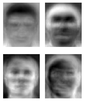
The above images are eigenfaces … which are statistically distilled basic components of human faces … from which ANY human face can be reconstructed as a combination of the above basic components. It’s a great mathematical trick – particularly if you’re into the whole mass surveillance and electronic police state thing.
If you are more into the whole, helping people and medical care thing, check out the global consortia at ENIGMA who have been carrying out massive genetic and brain scanning studies – like this one involving 437,607 SNPs in 31,622 voxels in 731 subjects using their new method, vGeneWAS, to study Alzheimer’s Disease:
“We hypothesized that vGeneWAS would, in some situations, have greater power to detect associations than existing SNP-based methods. One such situation might be when a gene contains many loci with weak individual effects. In addition, we expected that vGeneWAS would have greater overall power than mass SNP-based methods, like vGWAS, because of the drastic reduction in the effective number of statistical tests performed.”
The vGeneWAS method relies on the calculation of “eigenSNPs” which are eigenvectors that describe a matrix of n subjects by m SNPs in an individual gene (an n-x-m matrix of 1’s,0’s,-1’s for aa, aA, AA genotypes). EigenSNPs are sort of like eigenfaces insofar as eigenSNPs (which are not actual SNPs) capture the majority of variance, or the basic essence of an individual gene … but seriously, you should read the original article ’cause every stats test I ever took totally punched me in the face.
In his undergraduate writings while a student at Harvard in the early 1900’s E. E. Cummings quipped that, “Japanese poetry is different from Western poetry in the same way as silence is different from a voice”. Isabelle Alfandary explores this theme in Cummings’ poetry in her essay, “Voice and Silence in E. E. Cummings’ Poetry“, giving some context to how the poet explored the meanings and consequences of voice and silence. Take for example, his poem “silence”
silence
.is
a
looking
bird:the
turn
ing;edge, of
life
(inquiry before snow
e.e. cummings
Lately, it seems that the brain imaging community is similarly beginning to explore the meanings and consequences of the brain when it speaks (activations whilst performing certain tasks) and when it rests quietly. As Cummings beautifully intuits the profoundness of silence and rest, I suppose he might have been intrigued by just how very much the human brain is doing when we are not speaking, reading, or engaged in a task. Indeed, a community of brain imagers seem to be finding that the brain at rest has quite a lot to say – moreso in people who carry certain forms of genetic variation (related posts here & here).
A paper by Perrson and colleagues “Altered deactivation in individuals with genetic risk for Alzheimer’s disease” [doi:10.1016/j.neuropsychologia.2008.01.026] asked individuals to do something rather ordinary – to pay attention to words – and later to then respond to the meaning of these words (a semantic categorization task). This simple endeavor, which, in many ways uses the very same thought processes as used when reading poetry, turns out to activate regions of the temporal lobe such as the hippocampus and other connected structures such as the posterior cingulate cortex. These brain regions are known to lose function over the course of life in some individuals and underlie their age-related difficulties in remembering names and recalling words, etc. Indeed, some have described Alzheimer’s disease as a tragic descent into a world of silence.
In their recordings of brain activity of subjects (60 healthy participants aged 49-79), the team noticed something extraordinary. They found that there were differences not in how much the brain activates during the task – but rather in how much the brain de-activates – when participants simply stare into a blank screen at a small point of visual fixation. The team reports that individuals who carry at least one copy of epsilon-4 alleles of the APOE gene showed less de-activation of their their brain (in at least 6 regions of the so-called default mode network) compared to individuals who do not carry genetic risk for Alzheimer’s disease. Thus the ability of the brain to rest – or transition in and out of the so-called default mode network – seems impaired in individuals who carry higher genetic risk.
So, I shall embrace the poetic wisdom of E. E. Cummings and focus on the gaps, empty spaces, the vastness around me, the silences, and learn to bring my brain to rest. And in so doing, perhaps avoid an elderly descent into silence.
Joseph LeDoux‘s book, “Synaptic Self: How Our Brains Become Who We Are” opens with his recounting of an incidental glance at a t-shirt, “I don’t know, so maybe I’m not” (a play on Descartes’ “cogito ergo sum“) that prompted him to explore how our brain encodes memory and how that leads to our sense of self. More vividly, Elizabeth Wurtzel, in “Prozac Nation” recounts, “Nothing in my life ever seemed to fade away or take its rightful place among the pantheon of experiences that constituted my eighteen years. It was all still with me, the storage space in my brain crammed with vivid memories, packed and piled like photographs and old dresses in my grandmother’s bureau. I wasn’t just the madwoman in the attic — I was the attic itself. The past was all over me, all under me, all inside me.” Both authors, like many others, have shared their personal reflections on the fact that – to put it far less eloquently than LeDoux and Wurtzl – “we” or “you” are encoded in your memories, which are “saved” in the form of synaptic connections that strengthen and weaken and morph through age and experience. Furthermore, such synaptic connections and the myriad biochemical machinery that constitute them, are forever modulated by mood, motivation and your pharmacological concoction du jour.
Well, given that my “self” or “who I think of as myself” or ” who I’m aware of at the moment writing this blog post” … you get the neuro-philosophical dilemma here … hangs ever so tenuously on the biochemical function of a bunch of tiny little proteins that make up my synaptic connections – perhaps I should get to know these little buggers a bit better.
OK, how about a gene known as kalirin – which is named after the multiple-handed Hindu goddess Kali whose name, coincidentally, means “force of time (kala)” and is today considered the goddess of time and change (whoa,very fitting for a memory gene huh?). The imaginative biochemists who dubbed kalirin recognized that the protein was multi-handed and able to interact with lots of other proteins. In biochemical terms, kalirin is known as a “guanine nucleotide exchange factor” – basically, just a helper protein who helps to activate someone known as a Rho GTPase (by helping to exchange the spent GDP for a new, energy-laden GTP) who can then use the GTP to induce changes in neuronal shape through effects on the actin cytoskeleton. Thus,kalirin, by performing its GDP-GTP exchange function, helps the actin cytoskeleton to grow. The video below, shows how the actin cytoskeleton grows and contracts – very dynamically – in dendrites that carry synaptic spines – whose connectivity is the very essence of “self”. Indeed, there is a lot of continuing action at the level of the synapse and its connection to other synapses, and kalirin is just one of many proteins that work in this dynamic, ever-changing biochemical reaction that makes up our synaptic connections.
In their paper”Kalirin regulates cortical spine morphogenesis and disease-related behavioral phenotypes” [doi: 10.1073/pnas.0904636106] Michael Cahill and colleagues put this biochemical model of kalirin to the test, by examining a mouse whose version of kalirin has been inactivated. Although the mice born with this inactivated form are able to live, eat and breed, they do have significantly less dense patterns of dendritic spines in layer V of the frontal cortex (not in the hippocampus however, even though kalirin is expressed there). Amazingly, the deficits in spine density could be rescued by subsequent over-expression of kalirin! Hmm, perhaps a kalirin medication in the future? Further behavior analyses revealed deficits in memory that are dependent on the frontal cortex (working memory) but not hippocampus (reference memory) which seems consistent with the synaptic spine density findings.
Lastly, the authors point out that human kalirin gene expression and variation has been associated with several neuro-psychiatric conditions such as schizophrenia, ADHD and Alzheimer’s Disease. All of these disorders are particularly cruel in the way they can deprive a person of their own self-perception, self-identity and dignity. It seems that kalirin is a goddess I plan on getting to know better. I hope she treats “me” well in the years to come.
Few genes have been studies as intensely as apolipoprotein E (APOE). In particular, one of its variants, the epsilon-4 allele has been especially scrutinized because it is correlated with an earlier onset (about 10 years earlier than average) of Alzheimer’s Disease. Among the many roles of APOE – its just a tiny cholesterol binding protein – are those as participant in synaptic plasticity, early neural development, damage-response and other processes – all of which share the need for the synthesis and movement of neuronal membranes (see the fluid mosaic model) and their component parts – such as cholesterol. Hence, whenever neural membranes are being synthesized (plasticity & development) or damaged (overstimulation and other sources of oxidative damage) the tiny APOE is there to help with its membrane stabilizing cholesterol molecule in hand. Over the course of a lifetime, routine damage to neuronal membranes adds up (particularly in the hippocampus where constant storage-recall memory functions place enormous demands on synaptic plasticity systems), and individuals (such as epsilon-4 carriers) may simply show more wear-and-tear because their version of APOE is not as optimal as the other forms (epsilon-2 and -3).
With this etiological model in mind, perhaps you would like to take better care of you cell membranes (much like your car mechanic implores to change your car’s spark plugs and oil to keep the engine clean on the inside). Moreover, perhaps you would like to do-so especially if you knew that your APOE system was less optimal than average. Indeed, results from the recent REVEAL study suggest that folks who are in their 50’s are not unduly distressed to make this genetic inquiry and find out their genotypic status at this APOE polymorphism – even though those who discovered that they were epsion-4 carriers reported more negative feelings, understandably. Still, with a number of education and intervention strategies available, an optimistic outlook can prevail.
Furthermore, there are ever newer diagnostic strategies that can improve the rather weak predictive power of the genetic test. For example, cognitive assessments that measure hippocampal-dependent aspects of memory or visual orienting have been shown to be valid predictors of subsequent dementia – even moreso in populations that carry the APOE epsilon-4 allele. Other forms of neuroimaging that directly measure the structure and function of the hippocampus also have tremendous sensitivity (here for a broad review of imaging-genetics of AD) and can, in principle, provide a more predictive view into one’s distant future.
On the very cutting edge of this imaging-genetic crystal ball technology, lies a recent paper entitled, “Distinct patterns of brain activity in young carriers of the APOE-e4 allele” by Fillippini and colleagues [doi: 10.1073/pnas.0811879106]. Here, the research team asks whether individuals in their late 20’s show structural/functional brain differences that are related to APOE genotype. They employ various forms of imaging analysis such as a comparison of brain activity when subjects were performing a novel vs. familiar memory task and also an analysis of so-called resting state networks – which reflect a form of temporal coherence (brain areas that oscillate in-sync with each other when subjects are lying still and doing nothing in the scanner). For the analysis of the memory task, the team found that APOEe4 carriers showed more activation in the hippocampus as well as other brain regions like the anterior midbrain and cerebellum. When the team analysed a particular resting state network – the default mode network – they found differences in the medial temporal lobe (containing head of the hippocampus and amygdala) as well as the medial prefronal cortex. According to the paper, none of these differences could be explained by differences in the structure or resting perfusion of the young-adult brains in the study.
Wow, these results seem to suggest that decades before any mild cognitive impairments are observable, there are already subtle differences in the physiology of the APOEe4 brain – all of which could be detected using the data obtained in 6 minutes of rest.6 minutes of rest and spit in a cup – what does the future hold?


![Reblog this post [with Zemanta]](https://i0.wp.com/img.zemanta.com/reblog_c.png)


 With this